Learn the human body in 3D, structure by structure.

Fremma Anatomy is a 3D atlas for medical students. Turn the body, peel it layer by layer, and select any structure to see its name in English, French and Latin.

Drag to turn

Explore the atlas

Start from the part of the body you are studying, or follow one system through the whole body. Every picture here is the atlas itself.

By body region

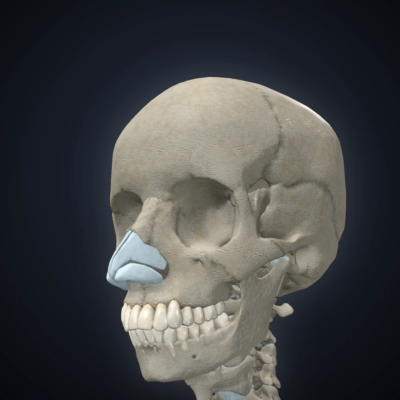
The skull in the atlas
HeadSkull, face and neck
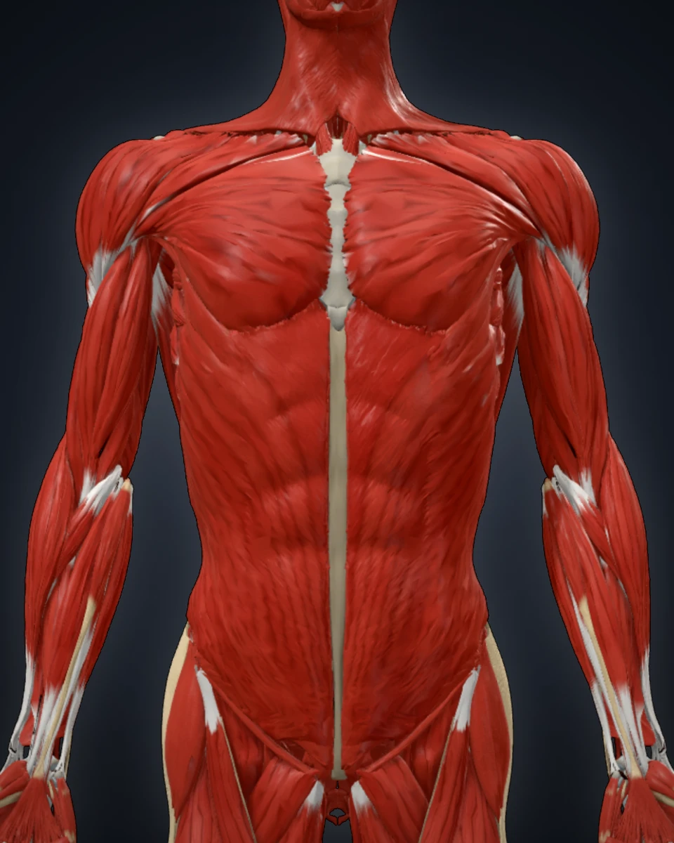
Muscles of the trunk in the atlas
TrunkThorax, abdomen and back
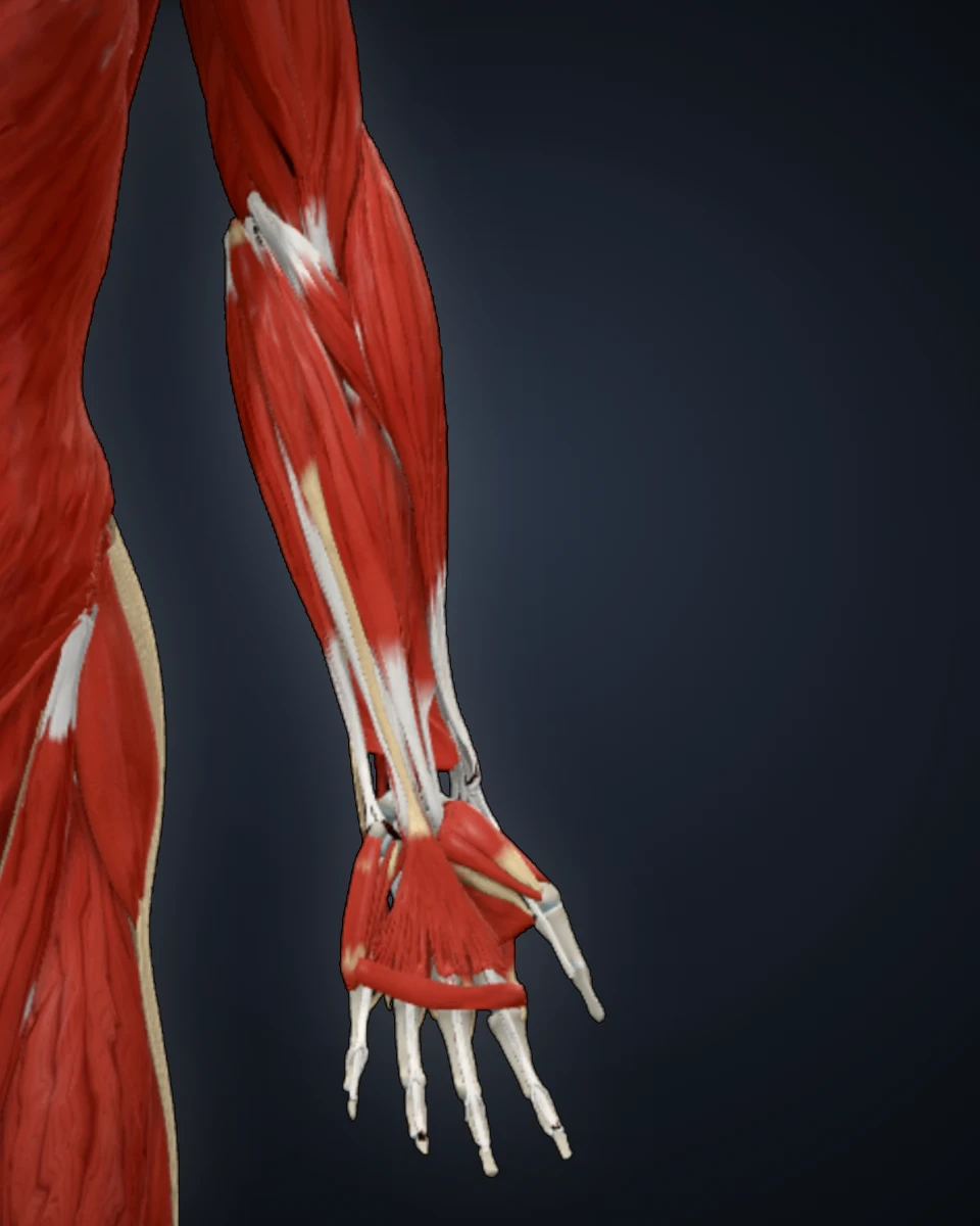
Muscles of the upper limb in the atlas
Upper limbShoulder to hand
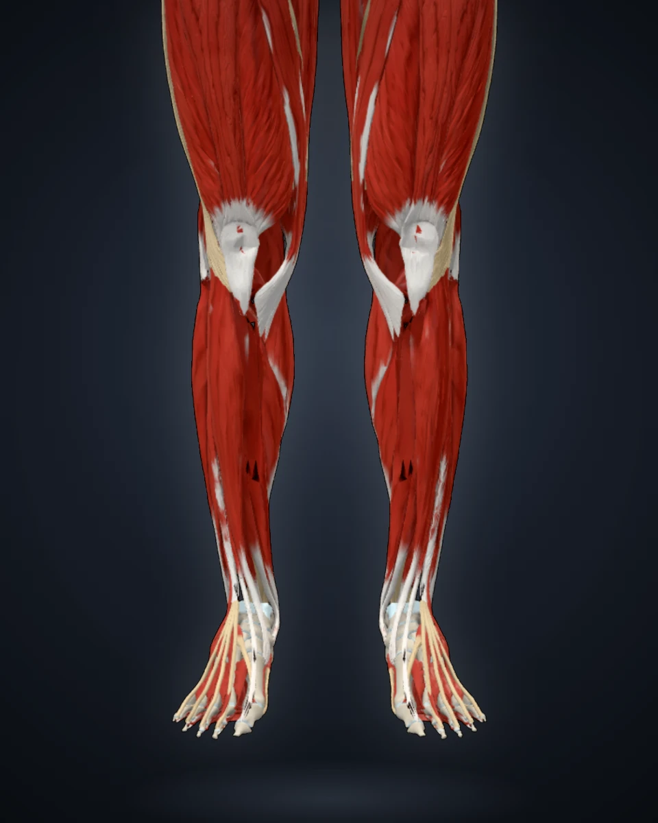
Muscles of the lower limb in the atlas
Lower limbHip to foot

By body system

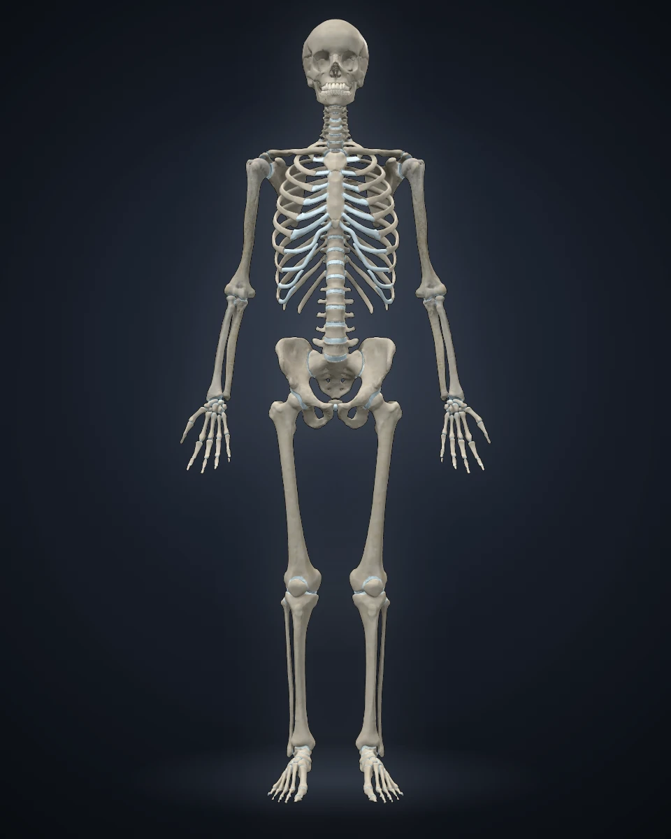
The skeleton in the atlas
Skeletal systemEvery bone of the body
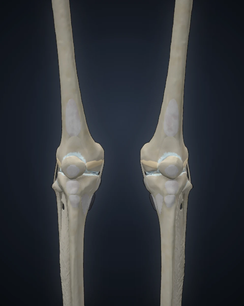
Joints and ligaments in the atlas
JointsCapsules, ligaments and discs
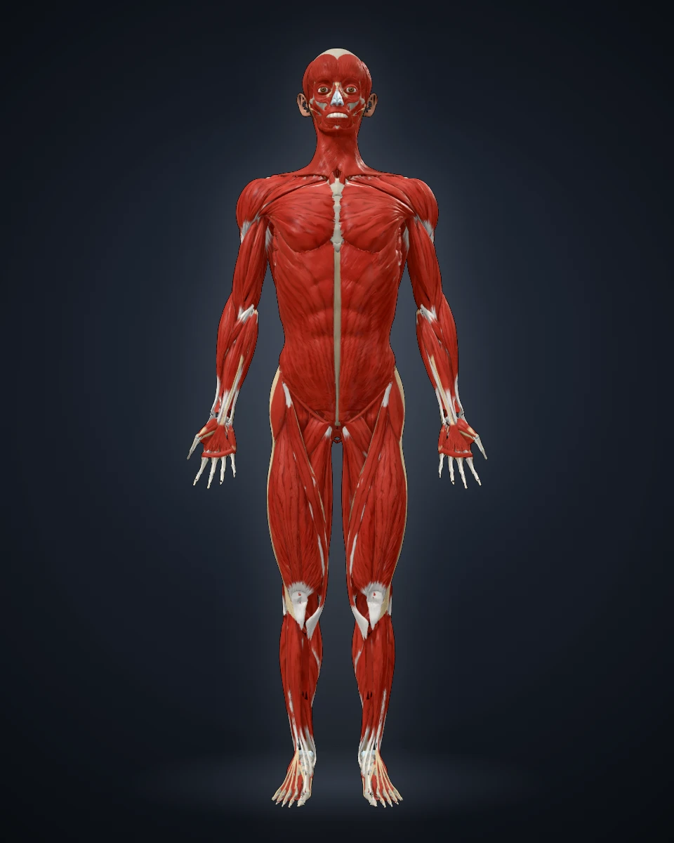
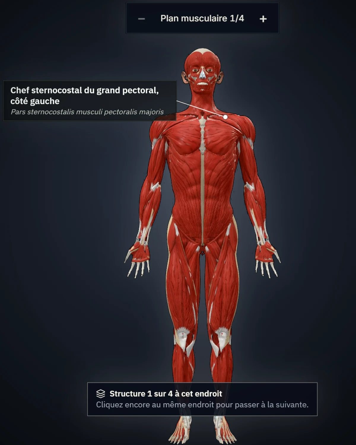
The muscular system in the atlas
Muscular systemMuscles and their attachments
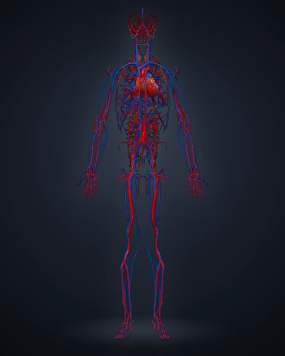
Heart, arteries and veins in the atlas
Cardiovascular systemHeart, arteries and veins
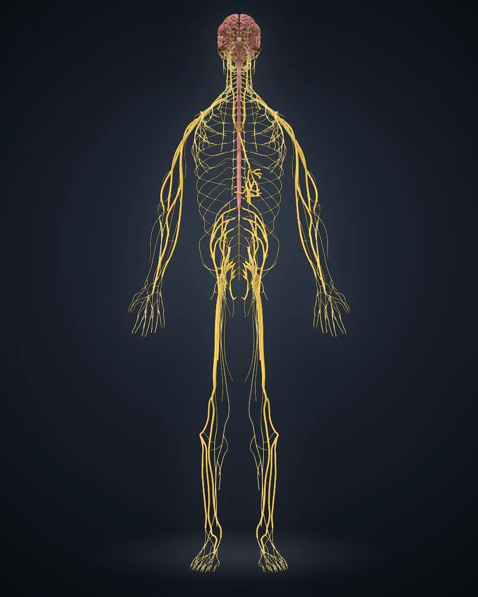
Brain, spinal cord and nerves in the atlas
Nervous systemBrain, spinal cord and nerves
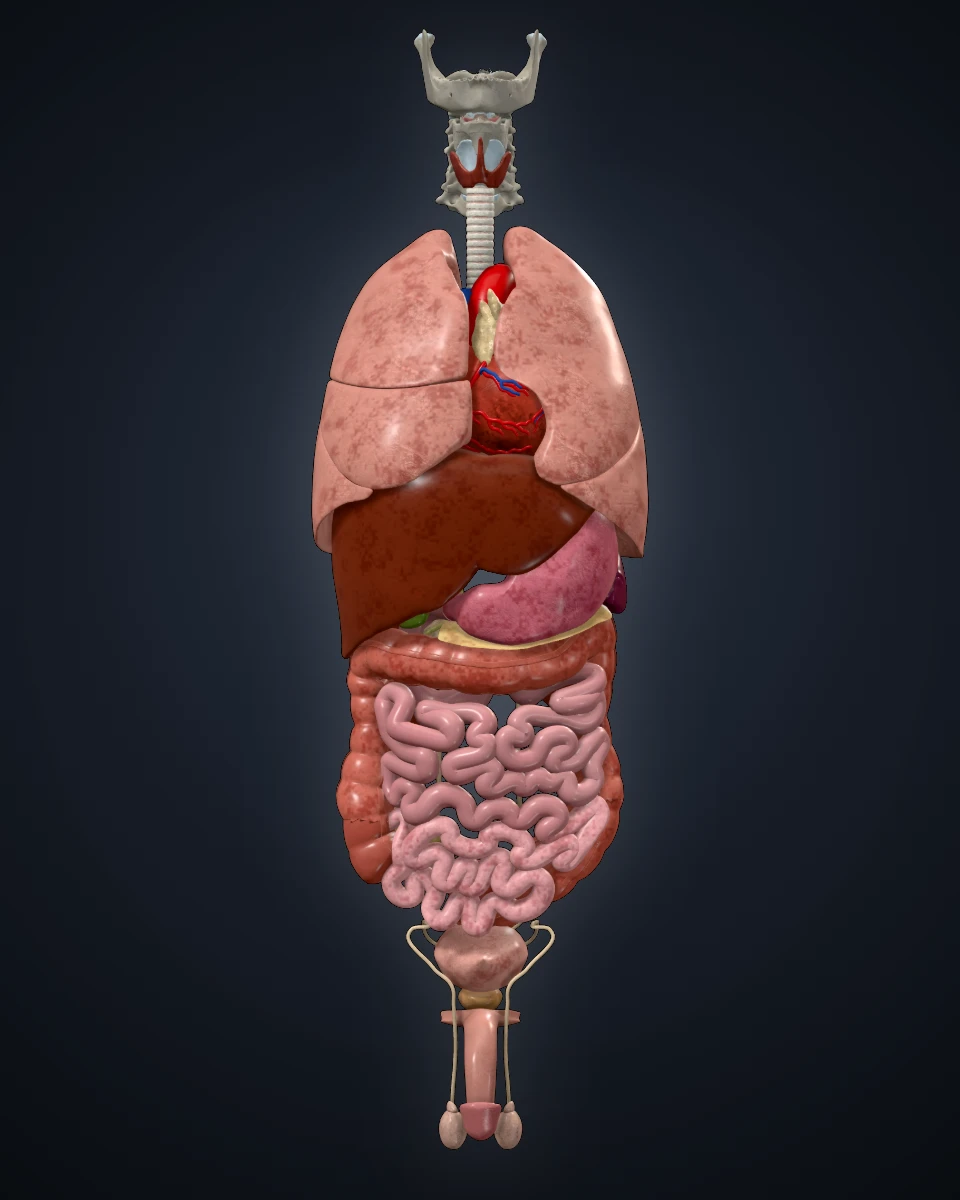
Thoracic, abdominal and pelvic organs in the atlas
OrgansChest, abdomen and pelvis

Inside the atlas

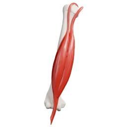
The atlas in use: the muscular system with one muscle selected and its names shown

Fremma Anatomy is made for medical students preparing their anatomy exams, and for the doctors who teach them. It sits beside the textbook: the book explains, the atlas shows the shape from every side.

A physician reviews each structure before students see it.

Names in three languages

English, French and the Latin term for every structure.

Available

Layer by layer

Bones, joints, muscles, vessels, nerves and organs, shown together or one at a time.

Available